Posts

Showing posts with the label UPSC

Explain the steps involved in retrograde transport

Retrograde transport is a process by which proteins and membranes are moved backward i.e., from later compartments to earlier ones within the endomembrane system. This transport mechanism plays an essential role in maintaining organelle identity, recycling transport machinery and retrieving specific proteins that need to return to their origin for reuse. It is important to understand from the beginning that retrograde transport is  not limited  to the retrieval of proteins from the Golgi apparatus back to the endoplasmic reticulum (ER). It also operates actively between endosomes and the Golgi, especially in the recycling of mannose-6-phosphate receptors (M6PRs). These receptors, after delivering lysosomal enzymes to endosomes, must return to the trans-Golgi network (TGN) to participate in another round of sorting. This ensures a continuous and efficient targeting of lysosomal enzymes. Retrograde transport is a multi-step process, usually involving four main steps, which toget...

Explain the steps involved in anterograde transport

Anterograde transport refers to the forward movement of proteins and lipids from the endoplasmic reticulum (ER) towards their final destinations, such as the Golgi apparatus, plasma membrane, or extracellular space. This process is a highly regulated and directional pathway, which ensures that proteins follow a proper route, allowing cells to maintain organisation, compartmentalisation and functional efficiency. The transport is carried out using vesicles, and involves several organelles and protein complexes working in coordination. This process can be described in five major steps, beginning in the rough ER and progressing toward the cell membrane or beyond: Step 1: Protein Synthesis and Folding in the Rough ER Anterograde transport begins with the synthesis of proteins on membrane-bound ribosomes of the rough ER. As the proteins are translated, they are inserted into the ER lumen or membrane. Within the ER, they undergo initial folding, assembly, and modifications, including N-linke...

Explain the targeting of soluble lysosomal proteins to endosomes and lysosomes

Image
The targeting of soluble lysosomal proteins, such as hydrolytic enzymes, to endosomes and lysosomes is a well-organised and highly regulated process. This mechanism ensures that such enzymes reach the lysosomes accurately, where they are needed for intracellular digestion, and are not secreted outside the cell. This entire process takes place through a series of interconnected steps, involving specific molecular signals, vesicle formation and organelle targeting, which begins in the rough endoplasmic reticulum (RER) and ends in the lysosome. Step 1: Synthesis in the Rough Endoplasmic Reticulum (RER) Soluble lysosomal enzymes are synthesised by ribosomes attached to the rough ER. As the mRNA is translated, the growing polypeptide is inserted into the ER lumen. Inside the ER, these proteins are properly folded and undergo N-linked glycosylation, which prepares them for further modifications in the Golgi. Step 2: Transfer to the Golgi Apparatus From the ER, the glycosylated proteins are t...

Explain the role of the ER and Golgi Complex in protein trafficking

Image
In eukaryotic cells, protein trafficking is a highly regulated and stepwise process that ensures proteins reach their correct destination either within the cell or outside of it. Two major organelles that perform central roles in this process are the  Endoplasmic Reticulum (ER) and the Golgi Complex.  Together, these organelles carry out multiple interconnected functions such as protein synthesis, folding, quality control, modification, sorting, and vesicle-mediated transport. Their roles are highly coordinated and form the backbone of the endomembrane trafficking system. Role of the Endoplasmic Reticulum (ER) The ER is the first organelle involved in the protein trafficking pathway. It exists in two forms, rough ER (RER) and smooth ER (SER). Among these, the rough ER is primarily involved in protein trafficking. It is called "rough" because it is studded with ribosomes, which are the sites of protein synthesis. The RER plays several important roles in protein trafficking: Sy...

Describe the endomembrane system

Image
The endomembrane system is a group of interconnected membrane-bound organelles found in eukaryotic cells. This system is responsible for the synthesis, modification, transport, and sorting of proteins and lipids within the cell. All organelles in this system are either physically connected or communicate with each other through vesicle-mediated transport. This system helps in maintaining the organisation, compartmentalisation and communication between different parts of the cell. The word "endomembrane" means "within the membrane" and this system is very important for keeping the cell functioning smoothly. Main Components of the Endomembrane System There are six major components that make up the endomembrane system. These are: 1. Nuclear Envelope The nuclear envelope is a double membrane that surrounds the nucleus. It contains nuclear pores that allow the controlled exchange of materials (like mRNA and proteins) between the nucleus and the cytoplasm. Its outer membr...

Describe the step involved in the trafficking of soluble lysosomal enzymes from the trans-Golgi network and cell surface to lysosomes

Image
The trafficking of soluble lysosomal enzymes from the trans-Golgi network (TGN) and sometimes the cell surface to the lysosomes is a highly regulated, multistep process. This mechanism ensures that lysosomal hydrolases are specifically recognized, sorted and delivered to their correct destination without getting misdirected or secreted. This sorting is based mainly on mannose-6-phosphate (M6P) tags present on the enzymes, which act as a signal for targeting. There are five major steps in this trafficking pathway: 1. Tagging with Mannose-6-Phosphate in the Golgi Apparatus Soluble lysosomal enzymes are first synthesized in the rough endoplasmic reticulum (RER) and transported to the cis-Golgi. Inside the Golgi, they are modified by a two-step enzymatic reaction: An enzyme called  N-acetylglucosamine-1-phosphotransferase  attaches a phosphorylated N-acetylglucosamine (GlcNAc-P) group to the mannose residue of the N-linked oligosaccharides on the enzyme. Another enzyme removes the...

What is the role of a mannose 6-phosphate residues in the sorting of protein?

Mannose-6-phosphate (M6P) residues play a critical role in the targeting and sorting of lysosomal enzymes from the trans-Golgi network to the lysosomes. This is one of the most well-known examples of protein sorting signals that help direct enzymes to their correct destination inside the cell. Lysosomes are cellular organelles responsible for breaking down macromolecules with the help of hydrolytic enzymes. These enzymes are synthesized in the rough endoplasmic reticulum (RER) and pass through the Golgi apparatus before reaching the lysosomes. However, they do not go to the lysosomes randomly. They are specifically tagged with mannose-6-phosphate residues, which act like an "address label" for lysosomal targeting. The process involves the following key steps: 1. Addition of Mannose-6-Phosphate in the Golgi: After synthesis in the RER, lysosomal enzymes enter the cis-Golgi, where they undergo post-translational modification. In the cis and medial Golgi, a special enzyme called...

Describe the types of coated vesicles and their functions

Coated vesicles are specialized membrane-bound carriers used for transporting proteins and lipids between different compartments inside the cell. They are called  "coated"  because their outer membrane is covered by specific protein coats that help in vesicle formation, cargo selection and targeting. There are three main types of coated vesicles: clathrin-coated, COPI-coated and COPII-coated vesicles. Each type is associated with a particular direction and function of transport. 1. Clathrin-Coated Vesicles Clathrin-coated vesicles are among the most extensively studied vesicles. These are mainly involved in transport between the trans-Golgi network, endosomes and the plasma membrane. Mechanism: Clathrin forms a unique structure made of three-legged units known as  triskelions,  which assemble into a lattice-like cage around the budding vesicle. This coat provides mechanical support and helps in vesicle formation. Clathrin works together with adaptor proteins, such as...

Describe the molecular mechanism of vasecular traffic

Vesicular traffic refers to the highly coordinated process by which proteins and lipids are transported between different compartments within the eukaryotic cell using membrane-bound vesicles. This mechanism is essential for maintaining compartmental identity, delivering newly synthesized proteins to their proper destinations, recycling membrane components and internalizing extracellular materials. The molecular mechanism of vesicular transport involves four key steps: vesicle formation, targeting, docking and fusion. 1. Vesicle Formation: The process begins at the donor membrane, where  cargo molecules  (proteins or lipids) are selected and packaged into transport vesicles. This step is mediated by coat proteins such as COPI, COPII and clathrin, which help in shaping the vesicle and selecting specific cargo. COPII-coated vesicles  transport proteins from the endoplasmic reticulum (ER) to the Golgi apparatus. COPI-coated vesicles  are involved in retrograde transport...

Differentiate between secretory and endocytic pathways of protein sorting

Image
Protein sorting in eukaryotic cells occurs through two major pathways,  the secretory pathway and the endocytic pathway.  Both are essential for maintaining intracellular organization, membrane composition, and regulated transport of proteins and other molecules. Both the secretory and endocytic pathways involve vesicular transport and share certain components but they differ fundamentally in their transport direction, functional role, origin and final destination within the cell. 1. Based on Definition and Direction of Transport The secretory pathway is a biosynthetic and outward pathway that transports newly synthesized proteins from the endoplasmic reticulum (ER) to the Golgi apparatus and finally to the plasma membrane or extracellular space. This pathway is responsible for the secretion of proteins and insertion of membrane proteins. In contrast, the endocytic pathway is an inward transport pathway where materials such as membrane proteins, fluids and macromolecules are i...

Explain the sorting of proteins to mitochondria

The sorting of proteins to mitochondria is an essential and complex process that ensures proteins synthesized in the cytoplasm reach their correct mitochondrial locations. Mitochondria have their own small genome, but most of their proteins are encoded by the nuclear genome. These nuclear-encoded proteins are synthesized in the cytoplasm and must be imported into the mitochondria, where they serve various functions, including energy production, metabolism and signaling. This import process involves several well-coordinated steps, including recognition, translocation and sorting to different mitochondrial compartments. 1. Synthesis and Recognition of Mitochondrial Targeting Signals The vast majority of mitochondrial proteins are encoded by the nuclear genome. These proteins are synthesized on cytosolic ribosomes and possess a specific targeting sequence known as the mitochondrial targeting signal (MTS), typically found at the N-terminal. This signal is usually a short hydrophobic sequen...

How are the nascent secretory proteins targeted to the ER?

The targeting of nascent secretory proteins to the endoplasmic reticulum (ER) is a highly coordinated and essential process in eukaryotic cells, especially for proteins that are destined to be secreted outside the cell or localized to membranes and organelles of the endomembrane system. This entire process occurs co-translationally, meaning that the protein is directed to the ER while it is still being synthesized by the ribosome. This mechanism follows a specific pathway called the  Signal Hypothesis,  first proposed by Gunter Blobel, which explains how newly forming proteins are recognized and directed to the ER membrane. The process includes five essential steps, which are explained in detail below: 1. Signal Sequence Recognition Nascent secretory proteins contain a special amino acid stretch called the signal peptide or signal sequence at their N-terminal end. This signal sequence is typically 15–30 amino acids long and contains a hydrophobic core that is recognized as the...

How translocation of secretory proteins takes place across the ER membrane?

The translocation of secretory proteins across the endoplasmic reticulum (ER) membrane is a crucial step in the secretory pathway. This process ensures that newly made proteins enter the lumen of the rough ER, where they begin their journey toward secretion or membrane insertion. Secretory proteins are synthesized by ribosomes in the  cytosol.  However, those meant to enter the ER begin their synthesis with a special short sequence of amino acids called a signal peptide at their  N-terminal end.  This signal peptide acts as a tag, directing the ribosome to the ER. As soon as the signal peptide emerges from the ribosome, it is recognized by a complex known as the  Signal Recognition Particle (SRP).  The SRP binds to the ribosome and signal peptide, temporarily pausing protein synthesis. This pause is necessary to guide the ribosome to the ER membrane without the protein being fully synthesized in the cytosol. The SRP-ribosome complex then docks on the ER mem...

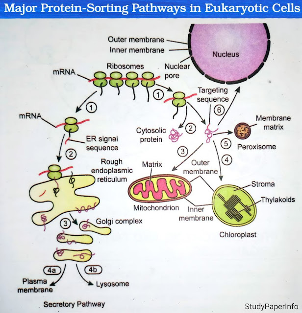
Explain with the help of schematic diagram, protein-sorting pathways in eukaryotic cells

Image
Protein sorting in eukaryotic cells is the highly regulated process through which newly synthesized proteins are directed to their correct destinations within or outside the cell. It is crucial for maintaining cellular structure, function and homeostasis. This process ensures that enzymes, structural proteins and signaling molecules reach their functional compartments such as the nucleus, mitochondria, endoplasmic reticulum (ER), lysosomes, plasma membrane, or extracellular space. The sorting mechanism relies on signal sequences in the proteins, cellular recognition systems and various sorting pathways. These pathways are crucial for maintaining cellular compartmentalization and function. There are two major types of protein-sorting pathways in eukaryotic cells: Co-translational targeting to the endoplasmic reticulum (ER) Post-translational targeting to other organelles like the nucleus, mitochondria and peroxisomes. 1. Co-translational Sorting Pathway (ER-Dependent Pathway) In this pa...

Desoribe endocytosis of LDL by the cell

Image
Low-Density Lipoprotein (LDL) is the primary transporter of cholesterol in the bloodstream. Cells acquire cholesterol from LDL particles through a highly specific process called  receptor-mediated endocytosis,  which is a classic example of clathrin-dependent endocytosis. This process begins when LDL particles bind to LDL receptors (LDLR) located on the  plasma membrane.  These receptors are transmembrane glycoproteins that specifically recognize apolipoprotein B-100 (ApoB-100) on the surface of LDL. The receptor-ligand complexes cluster in membrane regions called  clathrin-coated pits.  These regions are lined by the protein clathrin, which forms a polygonal lattice that shapes the invaginated membrane into a vesicle. As the clathrin-coated pit deepens, the membrane undergoes scission with the help of dynamin, a GTPase that pinches off the vesicle from the plasma membrane. This forms a clathrin-coated vesicle containing the LDL-receptor complexes. The clat...

What is endocytosis? Describe clathrin-independent and clathrin-dependent pathways of endocytosis

Endocytosis is a vital cellular process by which a cell engulfs extracellular materials by enclosing them in vesicles formed from the plasma membrane. It is an energy-dependent process (requiring ATP) that allows the internalization of nutrients, signaling molecules, fluids and even microorganisms. It helps maintain membrane composition, regulates receptor signaling and supports immune functions. This process is mainly of two types based on the presence or absence of the protein clathrin in vesicle formation. 1. Clathrin-Dependent Endocytosis Clathrin-dependent endocytosis is a highly selective and well-studied pathway where vesicles are formed with the help of a protein coat made of clathrin. The process begins when ligands (such as hormones or lipoproteins) bind to specific receptors on the cell membrane. These receptor-ligand complexes cluster together in specialized regions known as clathrin-coated pits. The clathrin lattice gives structural support and the pit invaginates, eventua...

Draw a schematic diagram to explain activation of postsynaptic receptors and cell response

Image
When a nerve signal travels across a synapse, the neurotransmitters released by the presynaptic neuron activate specific  postsynaptic receptors  on the membrane of the postsynaptic neuron or cell. This process is crucial for signal transmission between neurons and is responsible for various physiological responses like muscle contraction, memory formation and sensory perception. The activation of these receptors triggers a series of  cellular responses  that alter the electrical state of the postsynaptic neuron and determine whether it will generate an action potential. This process can be divided into two main parts: Activation of Postsynaptic Receptors:  This part includes the steps that happen when the neurotransmitters bind to the postsynaptic receptors and initiate a signaling process in the postsynaptic cell. Cellular Response:  This describes how the postsynaptic cell reacts after receptor activation, including changes in the membrane potential and ...

Describe the phases of cell excitation

Cell excitation mainly refers to the process through which excitable cells such as neurons and muscle cells respond to a stimulus by generating an electrical signal called an  action potential.  This signal helps in the transmission of information in neurons or in muscle contraction. The process of cell excitation occurs in well-defined electrical phases which are caused by controlled movement of ions (mainly Na⁺, K⁺, and sometimes Ca²⁺) across the cell membrane. These phases occur one after another in a sequence and each phase has a specific ionic mechanism and functional role. There are five main phases involved in the process of cell excitation: Resting Membrane Potential Depolarization Repolarization Hyperpolarization Restoration of the Resting Potential 1. Resting Membrane Potential The first phase in cell excitation is the resting membrane potential. At this stage, the cell is at rest and its membrane potential is stable, typically around -70mV, although this can vary sl...

What are the roles of leak channel, chemically gated channel, voltage gated channel and mechanically gated channel in cell excitation?

Cell excitation refers to the ability of a cell, especially a neuron or muscle cell, to respond to a stimulus by generating an electrical signal. This process mainly depends on the movement of ions across the cell membrane, which occurs through special proteins called  ion channels.  These channels control how ions like sodium (Na⁺), potassium (K⁺), calcium (Ca²⁺) and chloride (Cl⁻) enter or exit the cell. There are four main types of ion channels involved in this process: Leak Channels (also known as Passive Channels) Chemically Gated Channels (also called Ligand-Gated) Voltage-Gated Channels Mechanically Gated Channels 1. Leak Channels Leak channels are always open and allow continuous passive movement of specific ions, mainly potassium ions (K⁺), down their concentration gradient. These channels do not open or close in response to any external stimulus but remain open to maintain ionic balance. Role in excitation: Leak channels mainly help in maintaining the resting membran...

Describe in detail the channels responsible for cellular excitation

Image
Cellular excitation refers to the process by which a cell, especially nerve and muscle cells, becomes active and generates an  action potential.  This action potential is a rapid change in the membrane potential that allows the cell to send electrical signals over long distances. The ability of a cell to become excited and generate signals mainly depends on the presence and function of certain ion channels present in the plasma membrane. This process depends on the movement of ions like sodium (Na⁺), potassium (K⁺), calcium (Ca²⁺) and chloride (Cl⁻) across the cell membrane. Specialized proteins called  ion channels  regulate this ion flow. Based on how they open and function, ion channels involved in excitation are classified into four major types: Leak Channels, Voltage-Gated Ion Channels, Ligand-Gated Ion Channels and Mechanically-Gated Ion Channels. 1. Leak Channels (Passive Channels) Leak channels are ion channels that are either always open or remain open most ...

Popular posts from this blog

Desoribe endocytosis of LDL by the cell

What is a Cell? What Are the Essential Characteristics of Cells?

Describe the vesicular transport mechanism

What is endocytosis? Describe clathrin-independent and clathrin-dependent pathways of endocytosis

Describe the pattern of intermediate filaments' intracellular arrangement

Describe the molecular mechanism of vasecular traffic

What is the role of a mannose 6-phosphate residues in the sorting of protein?

Write the name of neurotransmitter which act as neuromodulator as well as inhibitor of neurotransmitter

Describe the step involved in the trafficking of soluble lysosomal enzymes from the trans-Golgi network and cell surface to lysosomes

Which neurological disorders are linked to increase dopamine secretion?